Email Us:info@neweastbio.com
Call Us:(+1) 610-945-2007

Email Us:info@neweastbio.com
Call Us:(+1) 610-945-2007
Size:100 μl
Price:$ 349
Brand:NewEast  Place of Origin:USA
Immunogen:
| Cat.#: N261198 | ||||
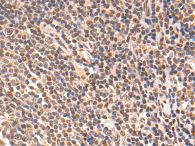
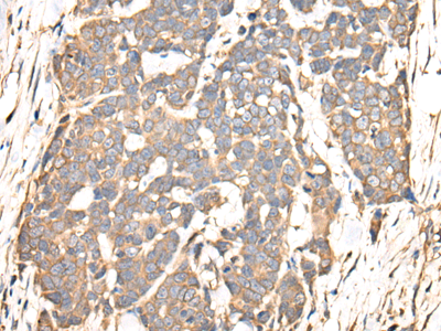
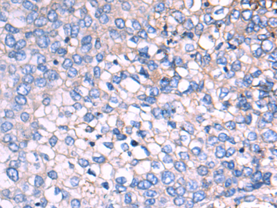
| Product Name: Anti-PINCH (3C12) Mouse Monoclonal Antibody | ||||
| Synonyms: LIMS1; PINCH; PINCH1; LIM and senescent cell antigen-like-containing domain protein 1; Particularly interesting new Cys-His protein 1; PINCH-1; Renal carcinoma antigen NY-REN-48 | ||||
| UNIPROT ID: P48059 | ||||
| Background: The protein encoded by this gene is an adaptor protein which contains five LIM domains, or double zinc fingers. The protein is likely involved in integrin signaling through its LIM domain-mediated interaction with integrin-linked kinase, found in focal adhesion plaques. It is also thought to act as a bridge linking integrin-linked kinase to NCK adaptor protein 2, which is involved in growth factor receptor kinase signaling pathways. Its localization to the periphery of spreading cells also suggests that this protein may play a role in integrin-mediated cell adhesion or spreading. Several transcript variants encoding different isoforms have been found for this gene. | ||||
| Immunogen: Purified recombinant fragment of human PINCH expressed in E. Coli. | ||||
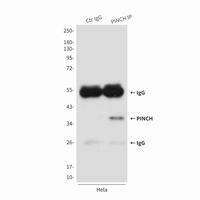
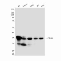
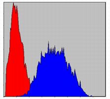
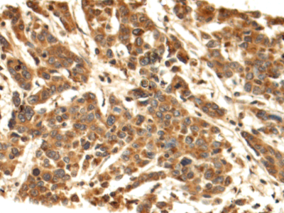
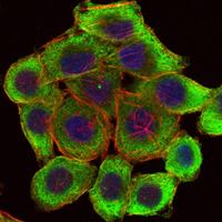
| Applications: WB,ICC/IF,FC,IP | ||||
| Recommended Dilutions: WB: 1/500-1/1000 IF: 1/50-1/200 IP: 1/20 FC: 1/50-1/100 | ||||
| Host Species: Mouse | ||||
| Clonality: Mouse Monoclonal | ||||
| Clone ID: 3C12-F7-A8 | ||||
| MW: Calculated MW: 37 kDa; Observed MW: 37 kDa | ||||
| Isotype: IgG1 | ||||
| Purification: Ascitic Fluid | ||||
| Species Reactivity: Human | ||||
| Conjugation: Unconjugated | ||||
| Modification: Unmodified | ||||
| Constituents: PBS (without Mg2+ and Ca2+), pH 7.3 containing 50% glycerol, 0.5% BSA and 0.02% sodium azide | ||||
| Research Areas: Cardiovascular | ||||
| Storage & Shipping: Store at -20°C. Avoid repeated freezing and thawing | ||||
|
Select By Alphabet
A B C D E F G H I J K L M N O P Q R S T U V W X Y Z
Subscribe to our latest email
(+1) 610-945-2007 info@neweastbio.com sale@neweastbio.com 840 First Avenue, Suite 400, King of Prussia, PA 19406
Copyright © 2010 - 2024 NewEast Biosciences | All rights reserved
Bioactive Transmembrane Proteins Antibodies for Transmembrane Proteins G Protein | GTPase


 Add to cart
 Download
